
THE HUMAN EYE
Regular exercise, a well balanced diet, a lot of green leafy vegetables, UV proection, and not smoking are keys to healthy eyes. The same blood vessels that run through our body are the same blood vessels transporting blood to and through our eyes. We always like to say “a healthy body = healthy eyes.” And never forget to wear 100% UV protection sunglasses when you’re spending time outdoors. The sun can easily damage our skin, so imagine what decades of sun exposure can do to the outside and inside your eyes.

THe CORNEA
The transparent outer cover of the eye that protects, the iris, anterior chamber and lens. The cornea is divided into 5 layers and is responsible for approximately two-thirds of the eye’s total optical power. It’s where we gently (hopefully) place our contact lenses when inserting them into our eyes. Without a healthy cornea what we see on a daily basis would be blurred and out of focus, so please, replace your contacts when you should, don’t abuse or sleep in them, and as always wear protective eye wear whenever it’s recommended or required.
THE iris
Or the “colored” part of our eye. It contains a group of muscles that are responsible for changing the diameter of the pupil to control the amount of light that enters our eyes. Think, low-light = large pupil; Bright light = small pupil. Damage to the iris via trauma can cause glaucoma. Conditions such as rheumatoid arthritis, ulceritive colitis, and IBS can cause the iris to be inflamed which causes your average eye to be extremely light sensitive and painful.


The Crystalline lens
This structure sits directly behind the iris and is responsible for the other one-third of the total optical power of the eye. It’s best known for becoming yellow and hazy over time and forming a cataract. Cataracts are very common in individuals 55+ and are easily removed by an opthalmologist and replaced with a clear artificial lens implant.

The retina is the innermost, light-sensitive layer of tissue of the eye of most vertebrates. Light striking the retina initiates a cascade of chemical and electrical events that ultimately trigger nerve impulses that are sent to various visual centers of the brain through the fibers of the optic nerve. Neural signals from the rods and cones undergo processing by other neurons, whose output takes the form of action potentials in retinal ganglion cells whose axons form the optic nerve. The macula (the dark red circular area in the photo above) is the area of the retina that is responsible for our central, fine detail vision.
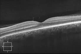
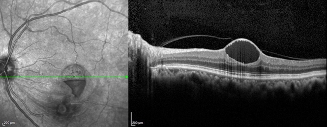
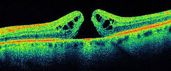
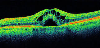
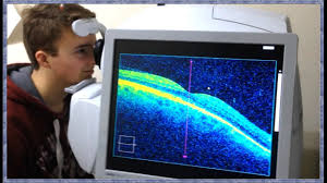
An OCT (optical coherence topography) scan pictured above and below, allows eye care providers to visualize the layers of the retina individually. These are commonly performed on individuals with macular degeneration or diabetic eye disease.

The optic nerve, also known as cranial nerve II, is the cable connection that transmits information from our retina, to our visual cortex via ganglion cells and glial cells. Damage to the optic nerve causing a progressive and irreversible loss of peripheral vision is known as glaucoma. Typically individuals with glaucoma have elevated eye pressures, but it’s possible to have normal eye pressures and have glaucoma. The shape of the optic nerve structure is similar to a bagel. The larger hole in your ‘bagel’, the higher your risk is for developing glaucoma. Early damage from glaucoma is almost always undetectable and asymptomatic.